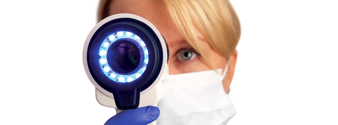
The VELscope® Vx, the latest model release of VELscope technology, uses natural tissue fluorescence to discover abnormalities in the oral mucosa.
The VELscope® Vx system enhances the way practitioners examine the oral mucosa and screen for tissue abnormalities, potentially leading to the earlier discovery of precancer, cancer or other disease processes. Early detection is one of the best mechanisms for enabling treatment success, increasing survival rates and maintaining a high quality of life.
The World’s #1 Adjunctive Examination Device
Traditional oral mucosal examination tools rely on reflected light to visualize the oral cavity. VELscope® Vx uses tissue fluorescence rather than reflectance. Natural tissue fluorescence is caused by “fluorophores” that, when excited by light of an appropriate wavelength (e.g. blue), will emit their own light at a longer wavelength (e.g. green). The resulting fluorescence can reveal a great deal about cellular, structural, and/or metabolic activity changes that are often directly related to disease processes occurring inside the tissue.
The VELscope® Vx system is a powerful device for the discovery of mucosal abnormalities such as:
- Viral, fungal and bacterial infections
- Inflammation from a variety of causes (lichen planus and lichenoid reactions, allergy to amalgam fillings, etc.)
- Squamous papillomas
- Salivary gland tumors
- Cancer and pre-cancer
- Other oral conditions
Unlike other adjunctive devices used by dentists, the VELscope® Vx system does not require any dyes or prolonged testing procedures. In fact, VELscope® Vx examinations can be performed in our office during routine hygiene exams (and under normal lighting conditions) in about two minutes.
VELscope® Vx Advantages
- Fits in with your work flow and complements your intra and extra oral head and neck examination with minimal set-up and only nominal time added to the overall appointment.
- Helps you find things which may be hard to see otherwise i.e. offers an imaging modality which is extremely sensitive to tissue changes.
- Provides visual information that is bright and easy to observe within the typical lighting conditions in a dental operatory.
- Allows for straightforward photo documentation – camera solution is easy to integrate and bright tissue response allows you to easily acquire digital images to be used for patient records or sent to referral partners.
- Clinically proven for the discovery of mucosal abnormalities, and for use by specialists in helping establish lesion margins for surgical excision.
- A very important tool in the fight against oral cancer.
- Readily accepted by patients and office staff.
- Well-supported in terms of after sales support, extensive training material as well as clinical support.
- Affordable for any practice.
- An attractive source of on-going revenue for dental practitioners.
VELscope® Vx is also the global market leader in tissue fluorescence visualization; over 25 million VELscope® Vx exams have been performed in 23 countries.